Monthly Archives: September 2018

Paper by Joh on analysis of lattice light sheet imaging data now published online in MBoC

Congratulations to Joh and former lab member Daphne on their paper “4D cell biology: big data image analytics and lattice light-sheet imaging reveal dynamics of clathrin-mediated endocytosis in stem cell derived intestinal organoids“, now published online in Molecular biology of the cell.

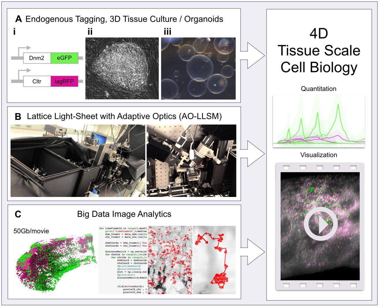
A new level of 4D tissue cell biology is unlocked as recent advances from three fields come together. (A) Endogenous protein tagging using genome editing (i), stem cell biology (ii) and 3D tissue/organoid culture (iii), (B) 4D non-invasive advanced fluorescent imaging with the lattice light-sheet microscope with adaptive optics (AO-LLSM, left: full view of the microscope, right: focus on the characteristic objective arrangement) and (C) advances and software in big data image analytics. (Right) Combination of these elements allows unprecedented quantitative analysis of subcellular events within live tissues in 4D.

Paper by Zane and Jonathan on microtubule dynamics accepted by JCS

Congratulations to Zane and Jonathan on their paper “Microtubule dynamics regulation reconstituted in budding yeast lysates“, now accepted and online in the Journal of Cell Science.

Recreation of Microtubule Dynamics in Budding Yeast Lysate. A reaction chamber is prepared by adhering GMPCPP-stabilized, rhodamine-labeled porcine microtubule seeds (red) to a glass coverslip through a biotin (black stems)-neutravidin (orange ovals) system. Whole-cell lysate from GFP-tubulin (green) expressing yeast strains is flowed into the chamber and incubated to allow for the polymerization of microtubules. Growth and dynamics of single microtubules is observed by TIRF microscopy in the context of other soluble proteins found in the cell (black shapes).