Paper by Michelle on a novel function of CDC42 published in JCB

Congratulations to Michelle on her paper “Cdc42 GTPase regulates ESCRTs in nuclear envelope sealing and ER remodeling ” now published in JCB.

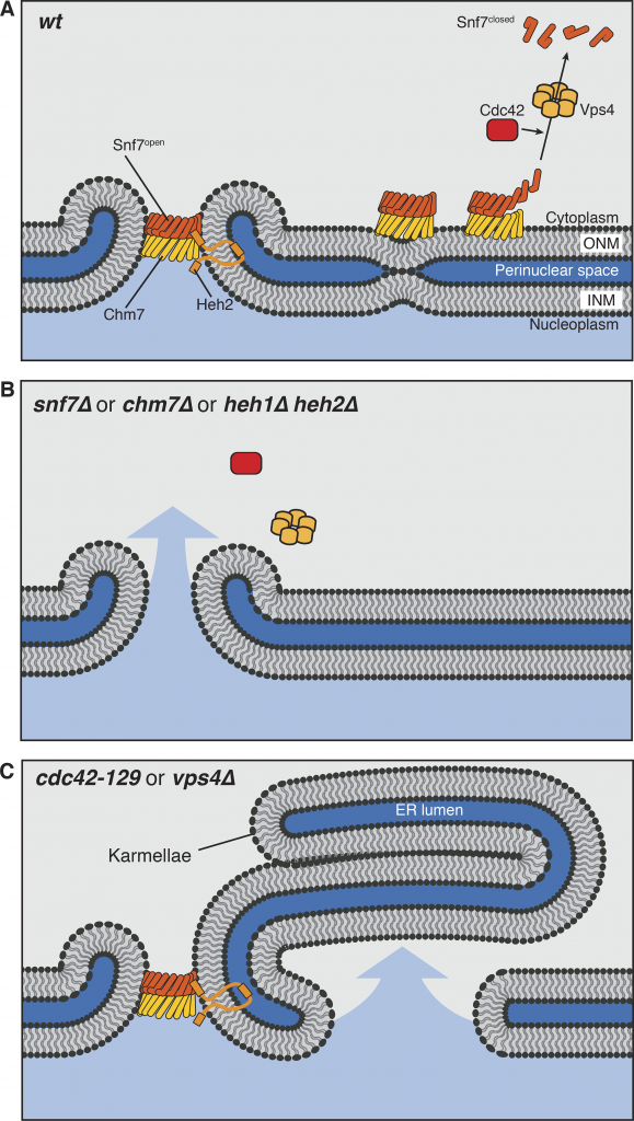
Cdc42, ESCRT-III, and Heh2 mutants share a leaky nucleus phenotype. (A) In normal cells, Heh2, Chm7, and Snf7 function at holes in the nuclear envelope to carry out annular fusion. ESCRT-III polymers are disassembled by Vps4, and we propose that Cdc42 is involved in the disassembly step by either directly contributing to Snf7 disassembly, activating Vps4 function, or cooperating with Vps4. (B) Mutants lacking components directly involved in annular fusion have holes in their nuclear envelopes left by nuclear fusion and ER fission events. (C) Cells lacking Vps4 and normal Cdc42 have unregulated ESCRT activity at the nuclear envelope. This causes the formation of nuclear karmallae and large holes in the nuclear envelope, leading to a defect in proper nucleo-cytoplasmic partitioning. ONM, outer nuclear membrane; INM, inner nuclear membrane.

Paper by Yidi Sun on budding vs. fission yeast CME now published in eLife

Congratulations to Yidi, her former undergraduate Tommy, Joh, Charlotte, and collaborators in the Xu Lab and Pollard Lab on their new paper “Direct comparison of clathrin-mediated endocytosis in budding and fission yeast reveals conserved and evolvable features” now published at eLife!

Comparison of endocytic vesicle formation in fission and budding yeast. Timeline and summary of the average molecule numbers for indicated coat proteins and actin machinery components in fission (A) and budding yeast (B) endocytosis. Scission occurs at the time 0. Steps (I) through (V) represent the full process from membrane invagination initiation to vesicle scission. See text (last section in Discussion) for detailed key similarities and differences in fission yeast and budding yeast in terms of protein numbers, architecture and function of the endocytic coat and associated actin machinery.

Paper by Ross Pedersen on functional role of endocytic myosins now published in JCB

Congratulations to Ross Pedersen on his new paper “Type I myosins anchor actin assembly to the plasma membrane during clathrin-mediated endocytosis“, now published in the Journal of Cell Biology.

Model for Myo5 function in anchoring actin assembly to the PM at endocytic sites. (A) Myo5 (yellow bananas) restricts activation of the Arp2/3 complex (gray avocados) by the WASP complex (blue widgets) to a discrete location, generating an actin array that grows predominantly in the same direction to generate force. (B) Absent this critical linkage, Arp2/3 activators splinter off of the PM, leading to Arp2/3 complex activation throughout the actin network. Delocalized Arp2/3 complex activation results in disordered actin arrays that fail to produce force. In the most catastrophic cases, the Arp2/3 complex and its activators pull away from the PM completely to form cytoplasmic actin comets (lower left of zoom).

Paper by Joh on analysis of lattice light sheet imaging data now published online in MBoC

Congratulations to Joh and former lab member Daphne on their paper “4D cell biology: big data image analytics and lattice light-sheet imaging reveal dynamics of clathrin-mediated endocytosis in stem cell derived intestinal organoids“, now published online in Molecular biology of the cell.

A new level of 4D tissue cell biology is unlocked as recent advances from three fields come together. (A) Endogenous protein tagging using genome editing (i), stem cell biology (ii) and 3D tissue/organoid culture (iii), (B) 4D non-invasive advanced fluorescent imaging with the lattice light-sheet microscope with adaptive optics (AO-LLSM, left: full view of the microscope, right: focus on the characteristic objective arrangement) and (C) advances and software in big data image analytics. (Right) Combination of these elements allows unprecedented quantitative analysis of subcellular events within live tissues in 4D.

Paper by Zane and Jonathan on microtubule dynamics accepted by JCS

Congratulations to Zane and Jonathan on their paper “Microtubule dynamics regulation reconstituted in budding yeast lysates“, now accepted and online in the Journal of Cell Science.

Recreation of Microtubule Dynamics in Budding Yeast Lysate. A reaction chamber is prepared by adhering GMPCPP-stabilized, rhodamine-labeled porcine microtubule seeds (red) to a glass coverslip through a biotin (black stems)-neutravidin (orange ovals) system. Whole-cell lysate from GFP-tubulin (green) expressing yeast strains is flowed into the chamber and incubated to allow for the polymerization of microtubules. Growth and dynamics of single microtubules is observed by TIRF microscopy in the context of other soluble proteins found in the cell (black shapes).

Goodbye Daphne, Itziar and Emily!

The Drubin/Barnes lab would like to send our fondest farewells to Daphne, Itziar and Emily who are moving on to exciting new positions!

Daphne Dambournet is now working as a Cell line engineer at Memphis Meats working to grow sustainable cultured meat.

Itziar Ibarlucea has taken a position as a post-doctoral associate in the laboratory of Jeffrey Ravetch at The Rockefeller University.

Emily Stoops is now working as a Scientist at Calico where she is working to combat aging.