Tag Archives: actin

Congratulations to Yansong Miao on his new paper!

Yansong Miao‘s new paper is out now at The Proceedings of the National Academy of Sciences.  Congratulations to Yansong on his great work!  The abstract is below.  The PDF can be downloaded from PNAS here.

Cell-cycle regulation of formin-mediated actin cable assembly. Miao Y, Wong CC, Mennella V, Michelot A, Agard DA, Holt LJ, Yates JR 3rd, Drubin DG. Proc Natl Acad Sci U S A. 2013 Nov 19;110(47):E4446-E4455. Epub 2013 Oct 16.  PMID: 24133141

Abstract

Assembly of appropriately oriented actin cables nucleated by formin proteins is necessary for many biological processes in diverse eukaryotes. However, compared with knowledge of how nucleation of dendritic actin filament arrays by the actin-related protein-2/3 complex is regulated, the in vivo regulatory mechanisms for actin cable formation are less clear. To gain insights into mechanisms for regulating actin cable assembly, we reconstituted the assembly process in vitro by introducing microspheres functionalized with the C terminus of the budding yeast formin Bni1 into extracts prepared from yeast cells at different cell-cycle stages. EM studies showed that unbranched actin filament bundles were reconstituted successfully in the yeast extracts. Only extracts enriched in the mitotic cyclin Clb2 were competent for actin cable assembly, and cyclin-dependent kinase 1 activity was indispensible. Cyclin-dependent kinase 1 activity also was found to regulate cable assembly in vivo. Here we present evidence that formin cell-cycle regulation is conserved in vertebrates. The use of the cable-reconstitution system to test roles for the key actin-binding proteins tropomyosin, capping protein, and cofilin provided important insights into assembly regulation. Furthermore, using mass spectrometry, we identified components of the actin cables formed in yeast extracts, providing the basis for comprehensive understanding of cable assembly and regulation.

Miao 2013 PNAS Figure 2

Average intensity projections along the z-axis of WT cells stained with Alexa-568 phalloidin to label actin filaments and expressing GFP-Tub1 as a cell-cycle stage indicator, obtained using 3D SIM microscopy.

Journal Club on Monday, August 6

For Journal Club on August 6, Eric Lewellyn presented the following paper:

Actin filament severing by cofilin dismantles actin patches and produces mother filaments for new patches. Chen Q, Pollard TD. Curr Biol. 2013 Jul 8;23(13):1154-62. PMID: 23727096.

Chen & Pollard Curr Biol 2013

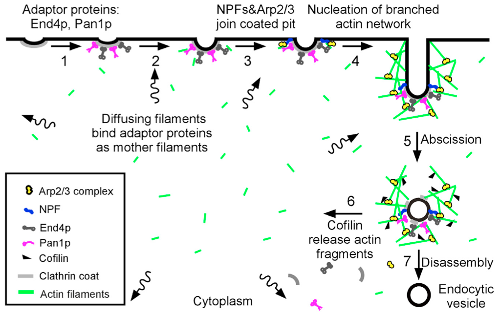
“Sever, diffuse and trigger” model for actin filament turnover in actin patches. The 7 steps (numbers next to the arrows) are (1) clathrin coated pits bind adaptor proteins End4p and Pan1p, (2) short, diffusing actin filaments bind to End4p and Pan1p associated with coated pits, and (3) Arp2/3 complex interacts with these mother filaments and nucleation promoting factors to (4) initiate branching nucleation of actin filaments that promote elongation of the endocytic tubule. (5) After abscission of the vesicle, (6) cofilin severs actin filaments to generate a pool of short, diffusing actin filaments, some of which return to the cycle at step 2.

Journal Club on Monday, March 17

For our Journal Club on March 17, Rebecca Lu will present the following paper:

Robust polarity establishment occurs via an endocytosis-based cortical corralling mechanism. Jose M, Tollis S, Nair D, Sibarita JB, McCusker D. J Cell Biol. 2013 Feb 18;200(4):407-18. PMID: 23401000

Schematics illustrating the mathematical model. Shown are the Cdc42 autoamplification module (A), the complete endocytosis module (B), the exocytosis module (C), and a legend for graphics (D).

Journal Club on Monday, Feb. 7

For our Journal Club on February 7, Yansong Miao will present the following paper:

Rocket launcher mechanism of collaborative actin assembly defined by single-molecule imaging. Breitsprecher D, Jaiswal R, Bombardier JP, Gould CJ, Gelles J, Goode BL. Science. 2012 Jun 1;336(6085):1164-8. doi: 10.1126/science.1218062. PMID: 22654058

Proposed ‘rocket launcher’ model for mDia1-APC collaboration during actin filament assembly

Congratulations to Yidi Sun on her new paper!

Yidi Sun‘s new paper is out now as an electronic publication ahead of print in the Journal of Cell Science.  Congratulations to Yidi on her great work!  The abstract is below.  The PDF can be downloaded from JCS here.

The functions of anionic phospholipids during clathrin-mediated endocytosis site initiation and vesicle formation. Sun Y, Drubin DG. J Cell Sci. 2012 Oct 24. PMID: 23097040.

Abstract

Anionic phospholipids PI(4,5)P(2) and phosphatidylserine (PS) are enriched in the cytosolic leaflet of the plasma membrane where endocytic sites form. In this study, we investigated the roles of PI(4,5)P(2) and PS in clathrin-mediated endocytosis (CME) site initiation and vesicle formation in Saccharomyces cerevisiae. Live-cell imaging of endocytic protein dynamics in an mss4(ts) mutant, which has severely reduced PI(4,5)P(2) levels, revealed that PI(4,5)P(2) is required for endocytic membrane invagination but is less important for endocytic site initiation. We also demonstrated that in various deletion mutants of genes encoding components of the Rcy1-Ypt31/32 GTPase pathway, endocytic proteins dynamically assemble not only on the plasma membrane but also on intracellular membrane compartments, which are likely derived from early endosomes. In rcy1Δ cells, fluorescent biosensors indicated that PI(4,5)P(2) only localized to the plasma membrane while PS localized to both the plasma membrane and intracellular membranes. Furthermore, we found that polarized endocytic patch establishment is defective in the PS-deficient cho1Δ mutant. We propose that PS is important for directing endocytic proteins to the plasma membrane and that PI(4,5)P(2) is required to facilitate endocytic membrane invagination.


Journal Club on Monday, October 15

For our Journal Club on October 15, Jay Ryoo presented the following paper:

The first five seconds in the life of a clathrin-coated pit. Cocucci E, Aguet F, Boulant S, Kirchhausen T. Cell. 2012 Aug 3;150(3):495-507. PMID: 22863004.

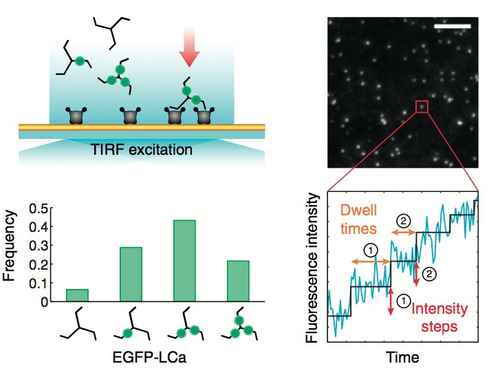
Schematic representation of the experimental setup and computational analysis

Journal Club on Monday, October 1

For our next Journal Club, Padmini Rangamani will present the following paper:

Phase transitions in the assembly of multivalent signalling proteins. Li P, Banjade S, Cheng HC, Kim S, Chen B, Guo L, Llaguno M, Hollingsworth JV, King DS, Banani SF, Russo PS, Jiang QX, Nixon BT, Rosen MK. Nature. 2012. PMID: 22398450

Phase transition correlates to biochemical activity transition in the nephrin/Nck/N-WASP system

Lighting Up Live Cells with Fluorescence (Genetic Engineering & Biotechnology News)

Genetic Engineering & Biotechnology News is out with a Feature Article this week including some comments from David Drubin about targeted genome modification in mammalian cells for fluorescence microscopy studies.

Lighting Up Live Cells with Fluorescence. Christine Herman. GEN. Sep 1, 2012 (Vol. 15, No. 32)

“The difference between taking snapshots of the process and watching a movie is just night and day,” says David Drubin, Ph.D., professor of cell and developmental biology at the University of California, Berkeley, whose lab uses fluorescence to understand the intricate details underlying clathrin-mediated endocytosis.


Researchers in David Drubin’s lab at the University of California, Berkeley genetically engineered a human cell line to express endogenous levels of RFP-tagged clathrin light chain A (red) and GFP-tagged dynamin 2 (green) for studying clathrin-mediated endocytosis. The above 3D kymograph of the cell surface, with the time dimension in the z-axis, shows the full lifetime of hundreds of clathrin patches on the membrane, which terminate upon recruitment of dynamin. [Aaron T. Cheng]

An Interview with David Drubin in Biowire

The May 2012 edition of Biowire, a publication of Sigma-Aldrich, includes an interview with David Drubin about the projects in our lab looking at clathrin-mediated endocytosis (CME) in mammalian cells using zing finger nuclease (ZFN) technology to undertake targeted genome modification. Traditionally, CME has been studied in cells in which fluorescently-tagged components of endocytic machinery are overexpressed using exogenous constructs. Data obtained in many labs using these methods suggested that CME was highly variable. Using ZFN technology, in collaboration with Sangamo Biosciences, our lab recently showed that CME is robust and efficient in mammalian cells.  The new results highlight the technical advantages of tagging genes at their endogenous loci, an approach that has been historically limited to genetically tractable organisms, such as the Drubin/Barnes Lab favorite Saccharomyces cerevisiae (budding yeast).  Emerging technologies, such as ZFNs and TALENs, however, are now making this sort of precise genomic manipulation possible in animal cells, including human cells, giving us new and powerful ways of studying cellular biology.

Cellular processes should be studied as close to their natural states as possible. I suspect that, as we see more uses of zinc finger nucleases [for tagging endogenous genes], people will find that they have been inadvertently perturbing the processes that they have been studying.

David Drubin (Biowire, May 2012)

Prof. David Drubin, for Biowire

Journal Club on Monday, June 11

For our next Journal Club, Lillie Cohn will present the following paper:

Reconstitution of clathrin-coated bud and vesicle formation with minimal components.  Dannhauser PN, Ungewickell EJ.  Nat Cell Biol. 2012 Apr 22;14(6):634-9.  PMID: 22522172.

Ultrathin section of H6-ΔANTH-AP180328–896-coated liposomes that were incubated with clathrin at 4 °C (c) or 37 °C